3.1
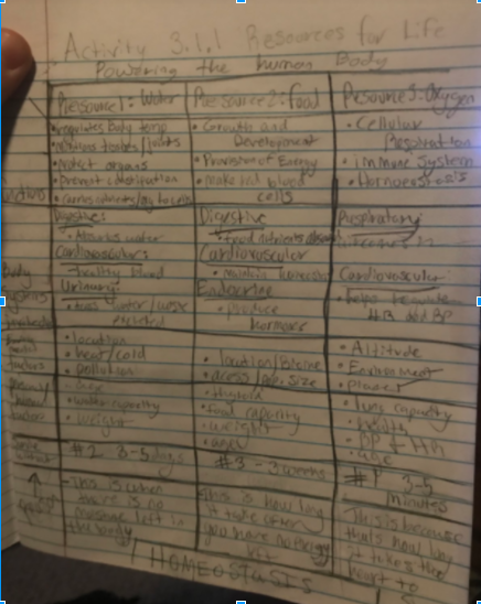
Activity 3.1.1 & 3.1.2 - Picture of Table: In this activity we looked at the resources our body needs to operate. In the table, it shows haw fast we need these 3 resources: food, water, and oxygen. This is where the Rule of 3´s come in.
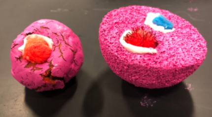
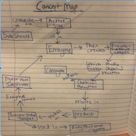
.Activity 3.2.1- Concept Map and Picture Constructed Product. The concept map shows how an enzyme acts on a substrate. In the constructed product, it shows what and enzyme attached to a substrate looks like.
|
|
3.2
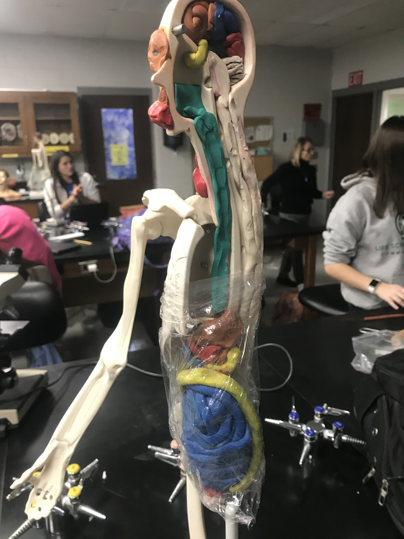
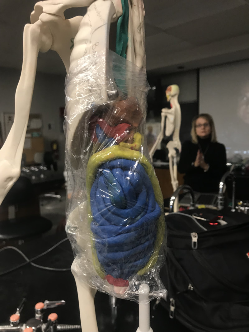
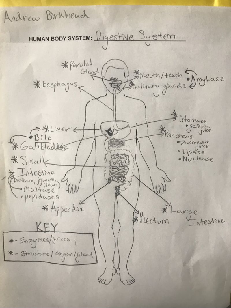
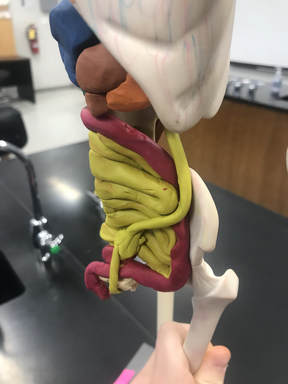
Activity 3.2.2- Body Organizer and Picture of Digestion System on the Manikin. This part of the Model shows the Digestive System of a human body. The humans Small intestine is 22 feet long!
|
|
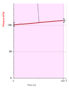
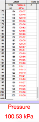
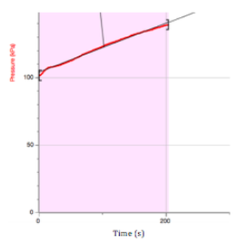
Activity 3.2.4- Pictures of Catalase Graphs and PowerPoint Presentation. In this activity, we found the effect enzyme concentration has on the chemical reaction. We concluded, the more Enzyme Concentration, the more reaction.
|
|
|
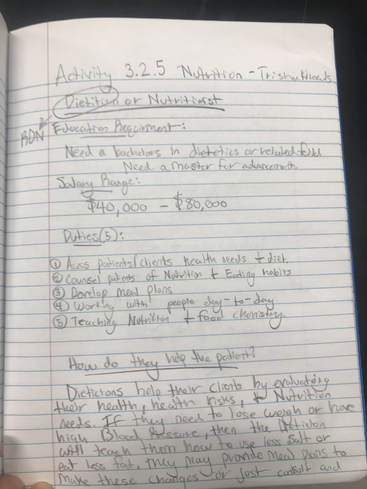
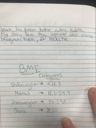
Activity 3.2.5- Client Report Documents and Career Journal. This picture show an outline of the job of a Dietitian. Also there is the categories of BMI shown in the second image.
|
|
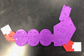
Activity 3.2.6- Picture of ATP Puzzle. This diagram made by kynsley and myself show the chemical make up of the ATP molecule. Which contains a phosphate group(which is where the energy come from), a base(adenine), and a ribose.
3.3
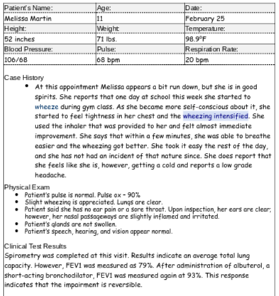
Activity 3.3.1 Visit #1 & #2 Resource Sheet. This the patients chart from her visit on February 5th. After a full workup and close examination for a few weeks; we confirmed the diagnosis of asthma.
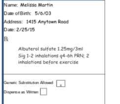
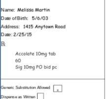
Activity 3.3.2 Visit #3 and Logger Pro Lung Capacity. This is Melissa third visit and her prescriptions give to her. The albuterol is the Rescue and the Accolate in the control.
|
|
|
Activity 3.3.3 Understanding Prescription Sheet
3.4
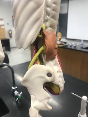
Activity 3.4.1 Manikin Picture. This part of our model represents the Renal System of the human body. As you can see, there is blood flow to the kidneys, the kidneys, ureters, bladder, and a urethra.
|
|
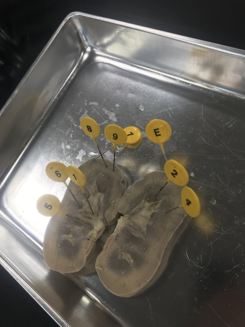
Activity 3.4.2 Dissected Kidney. The picture shows a perfectly sliced in 1/2 kidney with all its main parts. It is the kidney of a pig. Below is the key for the numbers on the kidney.
- Renal Pelvis 1
- Renal Medulla 2
- Renal Cortex E
- Medullary Pyramid 4
- Ureter 5
- Calyx 6
- Renal Capsule 7
- Site of the Glomerulus and Bowman’s capsule 8
- Site of Collecting Ducts and Loop of Henle 9
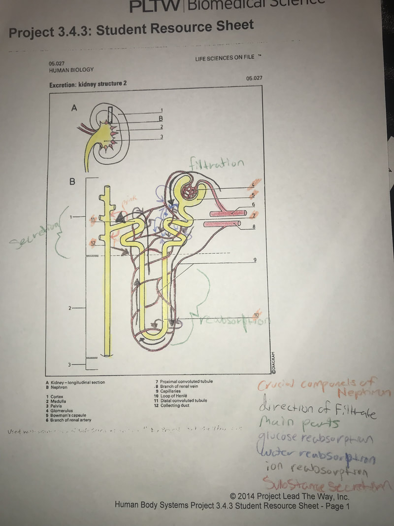
Activity 3.4.3 Nephron Diagram. Below is the handout for the section on the Nephron. There are about a million in each kidney. Its responsibilities are Filtration, Resorption, and Secretion.
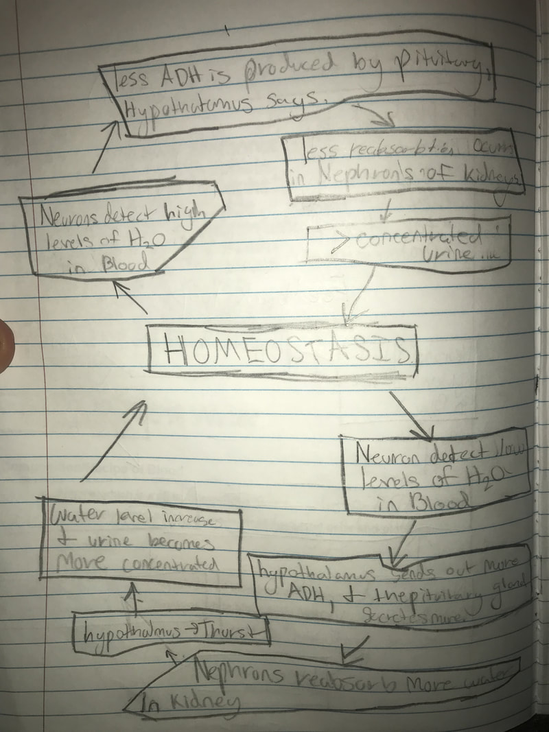
Activity 3.4.4 Inspiration Feedback Loop. This is my NEGATIVE feedback loop for the regulation of water in the body and the role ADH plays in that. There are many parts to this loop: Hypothalamus, Pituitary Gland, Urine, kidney, Ureters, water, ADH, thirst.
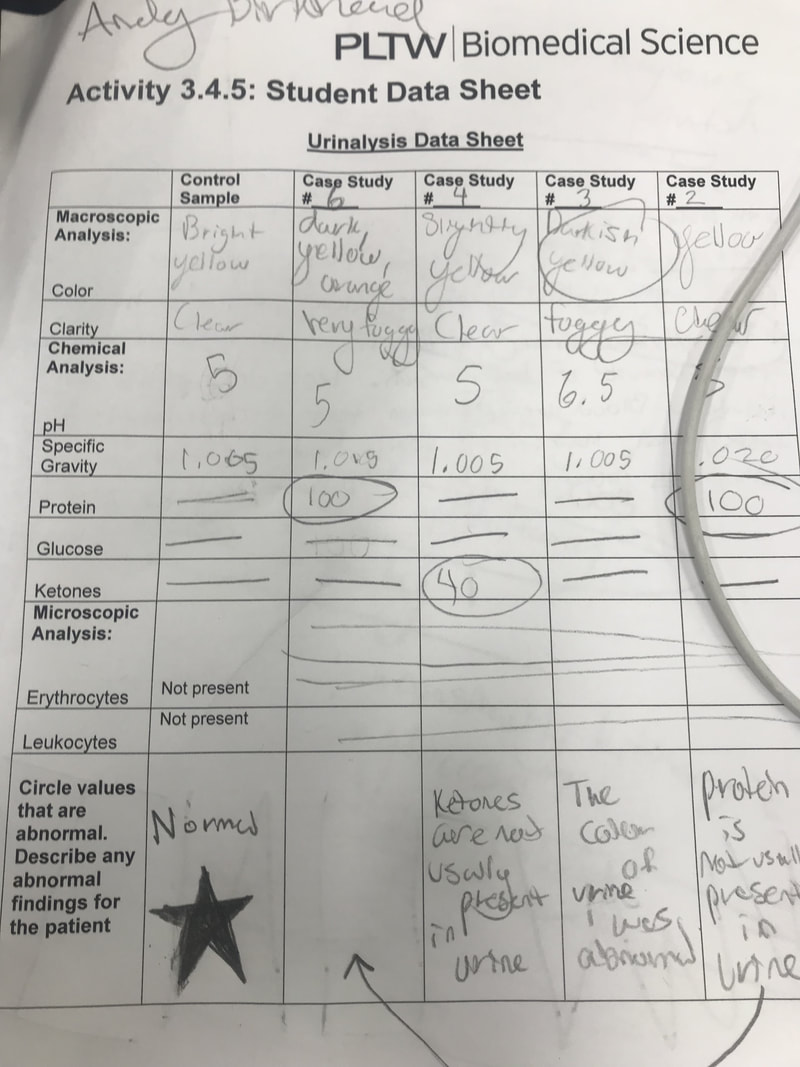
Activity 3.4.5 Urinalysis Student Data Sheet and PP presentation. In this activity we examined many different case study's and their urine. We examined the chemical composition of their urine as shown. Case study 2 was found to have a Exercise Induced protienuria.